
Page 48 - IJB-8-1
P. 48
Controlling Droplet Impact Velocity and Droplet Volume Improves Cell Viability in Droplet-Based Bioprinting
A
B
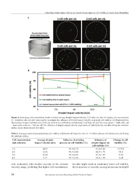
Figure 6. Increasing cell concentration leads to slower average droplet impact velocity. Cell-laden bio-inks of varying cell concentration
(1 – 4 million cells per mL) were used to investigate the influence of droplet impact velocity on printed cell viability. (A) Representative
fluorescence images of printed cells (20 nL per spot) on dry well-plates stained using Live/Dead cell viability assay (green – viable cells, red
– dead cells); scale bar = 200 µm. (B) The influence of droplet impact velocity on printed cell viability before and after hitting the substrate
surface on dry tissue-treated well plate.
Table 4. Average droplet impact and printed cell viability of different cell-laden bio-inks (1 – 4 million cells per mL) before and after hitting
the substrate surface.
Cell concentration Average droplet Influence of printing Influence of Change in cell
(mil cells/mL) impact velocity (m/s) process on cell viability (%) droplet impact on viability (%)
cell viability (%)
1.0 14.07 95.3±2.78 67.4±3.86 −27.9%
2.0 11.81 94.3±2.02 82.5±1.74 −11.8
3.0 10.52 93.1±2.63 87.3±1.97 −5.92
4.0 5.77 92.7±2.38 92.2±1.99 −0.44
only moderately with droplet viscosity in the relevant bio-inks might result in moderately lower cell viability,
viscosity range, predicting that higher cell concentration due to increase in viscosity causing an increase in droplet
34 International Journal of Bioprinting (2022)–Volume 8, Issue 1