
Page 135 - IJB-8-2
P. 135
Li, et al.
would quickly heal after microneedle treatment. The skin The insulin injection dose of two IU/kg was selected for
of mice was observed with the naked eye at 0, 10, 20, the positive control group and experimental group to
30, 60, and 120 min after the HMNP application. Pictures avoid hypoglycemia. The blood glucose levels were then
were taken with a camera for recording them. monitored at 0, 0.5, 1, 2, and 4 h after insulin injection.
In the negative control group, mice received no other
2.8. Preparation of HMN syringe treatment except that blood was collected at the same
A 3D-printed HMNP was pasted after post-treatment time to monitor blood glucose levels.
on the punched steel plate with quick-drying adhesive. 2.12. Ethics statement
The punched steel plate had the same size as HMNP,
and the holes in the plate correspond to the cavities of The experimental procedures were approved by the
microneedles. The HMN syringe was then formed by Institutional Animal Care and Use Committee of West
connecting HMNP at one end of the converter and China Hospital of Sichuan University (20211424A) and
a micro-syringe at the other end. The converter was were performed in accordance with the guidelines and
customized by 3D printing technology of SLA to match regulations of the Sichuan University Committee on
HMNPs and micro-syringes. Animal Research and Ethics.
2.9. Delivery ability of HMN syringe in vitro 2.13. Statistical/data analysis
The agarose gel was used as an in vitro skin model and All data were analyzed and arranged using GraphPad
rhodamine as the model drug to assess the in vitro drug Prism 8 (GraphPad Software Inc., CA, USA). The
delivery ability of the HMN syringe. According to the significance of the data was calculated using Student’s
literature , agarose powder was added with distilled t-test or one-way ANOVA methods. Results were
[27]
water (0.0265 g/mL), heated to 100°C until completely presented as means with standard error of the mean and a
dissolved, and then poured into the mold for cooling and P < 0.05 was considered statistically significant.
molding for later use. Rhodamine was slowly injected
into the above skin model using the HMN syringe, and 3. Results
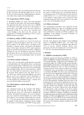
the delivery process was photographed under an optical 3.1. HMNPs customized by SOPL
microscope.
Schematic diagram for fabricating HMNPs via SOPL is
2.10. Mouse models of diabetes depicted in Figure 1. During the printing process, according
to the printing picture, a light beam was modulated into a
C57BL/6 mice were placed in a pathogen-free room with
a 12 h light-dark cycle. The diabetic mouse was induced customized pattern by a DMD and then passed through a
high magnification microlens, which significantly improved
by STZ. Generally, male C57BL/6 mice (6 – 8 weeks old) the printing resolution to 5 μm. An annulus consisting of
were intraperitoneally injected with 2% STZ (150 mg/kg) two non-concentric circles, namely a large white circle and
after overnight fasting. Mice were then fed with a normal a small black circle was designed in the printing picture
diet. One week later, the experiment was carried out in to customize the HMN. The white circle represented the
the morning where diabetic mice had fasted for 12 h. area exposed by the digital light, and the black circle was
The blood was collected from tail clipping, and blood the unexposed area. The formation principle of HMNs
glucose levels were monitored with a glucometer of was based on the spatial intensity distribution of projected
Yuwell. Fasting blood glucose higher than 16.7 mmol/L annulus light. On the one hand, the light intensity distribution
(or 300 mg/dl) were confirmed as type 1 diabetic mice of circle light gradually weakened from the center of the
and were used for further experiment .
[28]
focal plane to the outside and resembled an inverted cone.
2.11. Blood glucose control study in type 1 The small black circle represented nonexposed area, thus
diabetic mice forming a hollow-cone structure. On the other side, the
light intensity was greatly reduced after being absorbed
The experiment began in the morning with diabetic by photosensitive resin according to the Beer-Lambert
mice fasted for 12 h. The hair of mice was partially Law. The unique distribution of light intensity enabled the
shaved the day before the experiment to facilitate the formation of HMNs accordingly. In addition, we simulated
injection. The initial blood glucose levels of the diabetic the light propagation and light intensity distribution during
mice were measured at first. Diabetic mice with similar printing (see video in Supplementary File) to visualize the
blood glucose were randomly divided into three groups formation process of the HMN. It can be observed that the
(n = 5 for each group) with untreated diabetic mice as hollow-cone light intensity distribution was adjusted with
the negative control group, commercial insulin syringe the curing of the monomer solution, and finally, the HMN
as the positive control group, and HMN syringe group. was formed.
International Journal of Bioprinting (2022)–Volume 8, Issue 2 127