
Page 47 - IJB-8-2
P. 47
Jiao, et al.
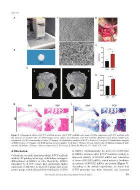
A C D
F
B
E
G
H I
Figure 5. Osteogenesis effects of β-TCP scaffolds in vivo. (A) β-TCP scaffolds were made. (B) The appearance of β-TCP scaffolds with
the diameter of around 7 mm. (C) SEM images of the surface microstructure of β-TCP scaffolds. (D) Rat cranial defect models were
constructed. (E) 3D reconstruction of micro-CT images. (F) Quantitative analysis of BV/TV of micro-CT images. (G) Quantitative analysis
of BMD of micro-CT images. (H) H&E staining of skull samples. Scale bars = 500 μm, 100 μm, respectively. (I) Masson staining of skull
samples. Scale bars = 500 μm, 100 μm, respectively. S: Soft tissue; B: Bone; M: Material. *P < 0.05; **P < 0.01
4. Discussion in BMSCs. Mechanistically, the m6A level of RUNX2
in BMSCs increased after β-TCP treatment, leading to
Collectively, our study demonstrated that β-TCP scaffolds
made by 3D printing technology could induce osteogenic improved stability of RUNX2 mRNA and retardation
differentiation of BMSCs in vitro. Meanwhile, BMSCs of decay of RUNX2 mRNA, which indirectly facilitates
stimulated by β-TCP extract had significantly higher an increase of RUNX2 mRNA and protein (Figure 7).
expression of METTL3 in the β-TCP group than in the According to the animal experiments, we found that
control group, which affected m6A modification of RNA β-TCP promoted new bone formation and increased
International Journal of Bioprinting (2022)–Volume 8, Issue 2 39