
Page 64 - IJB-8-3
P. 64
Extrusion of two-vasculature scaffold for angiogenesis
2.3. Two-vasculature-embedded scaffold immersed in culture media. The glass tube has a tapered
formation structure with about 1000 µm ID at one end and about
2000 µm ID at the other end. Using a syringe pump,
Three syringe pumps (11 Elite C300918, Harvard the 1000 µm outer diameter (OD) scaffold was sucked
Apparatus, U.S.A.) were connected to the fabricated and fixed into the narrow end of the tapered tube. The
device through Tygon tubes (Saint-Gobain, Courbevoie, scaffold was fixed in the tapered glass tube soaked in
France). For one core inlet, a mixture of 3 mg/mL culture media. To make conventional media diffusion
collagen, 2 × 10 cells/mL HUVECs, and 0.1 M CaCl condition into the embedded cells, the generated scaffold
6
2
(Daejung Chemicals, Republic of Korea) was supplied to was just soaked in culture media. To supply culture media
culture into a blood vessel. For another core inlet, 0.1 M through the hollow channel of the generated scaffold,
CaCl was injected to formulate a hollow channel inside
2
the scaffold. For the outer layer inlet, a 2% w/v mixture one end of the generated scaffold was sucked and fixed
of gelatin (Sigma-Aldrich, U.S.A.) and alginate (Daejung at the holder of the laboratory made connecting device.
Chemicals, Republic of Korea) (70 vs. 30 ratio) was Alginate was used to fill the gap between the holder and
supplied as the body of the scaffold. The extruded scaffold the fixed scaffold for a secure connection. Culture media
was submerged into a 0.1 M CaCl bath through the outlet were provided at a flow rate of 10 µL/min from a syringe
2
and then cross-linked. Calcium ions of the CaCl cross- pump to the connected scaffold and then flowed out the
2
linked with sodium alginate into calcium alginate so that not connected end of the scaffold. At the initial status of
no hydrogel in the hollow channel remained. The gelatin the media supply, there have been no culture media out
scaffold was washed with phosphate-buffered saline of the scaffold. However, as culture media flowed out
(PBS, Sigma-Aldrich, U.S.A.). The washed scaffold continuously, the dumped media made a puddle around
was cultured in an incubator at 37°C with 5% CO and the scaffold. The media puddle trashed out of the Petri
2
replaced with a fresh medium every 2 days. dish every 24 h.
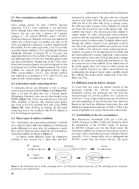
2.4. Laboratory made connecting device 2.6. Diffusion from the hollow channel
A connecting device was fabricated to link a syringe To select flow rate inside the hollow channel of the
pump to the generated scaffold (Figure 3 and Figure S1). generated scaffold, the cell-free two-vasculature-
First, a 2.0 mm ID glass tube and a Pasteur pipette embedded scaffold was produced, and red food dye
(Hilgenberg, Germany) were cut and then bonded using flowed through the cell-free scaffold from 2 µL/min to
PDMS (Figure 3A). A hole was punched at a Petri dish 20 µL/min. Using a bright-field microscope, the diffusion
(SPL, Republic of Korea). The attached glass holder rate and morphology of the red food dye were observed.
was fixed at the hole punched Petri dish using PDMS Based on the food dye diffusion observation, flow and
(Figure 3B). After checking no leakage, the fabricated diffusion of green fluorescence dye in PBS (1:1000) were
connecting device was sterilized with 99.9% ethanol in analyzed quantitatively using a fluorescence microscope.
24 h for biological experiments.
2.7. Perfusibility in the two vasculatures
2.5. Three types of culture condition Blue fluorescence microbeads (5.42 µm ± 0.09 µm,
The formulated two-vasculature-embedded scaffolds GmbH, Germany) in PBS (1:200) were flowed to check
were cultured in three different conditions: (i) Obstructing the perfusibility of the HUVEC vessel. To develop
media diffusion by a glass tube; (ii) soaking in a media the HUVEC-collagen core into the blood vessel, the
dish; and (iii) flowing media inside the two-vasculature- formulated scaffold was cultured in the soaking condition
embedded scaffold, as shown in Figure 4. for 2 days. After 2-day maturing, Calcein AM dye (Thermo
To hinder media diffusion inside the formulated Scientific, U.S.A.) stained alive cells to distinguish the
scaffold, it was inserted into a glass tube and then HUVEC vessel from the hollow channel. The green
A
B
Figure 3. The fabrication process of the connecting device. (A) The holder fabrication process. (B) The attaching process of the hole
punched Petri dish and the holder.
56 International Journal of Bioprinting (2022)–Volume 8, Issue 3