
Page 65 - IJB-8-3
P. 65
Nguyen, et al.
A B C
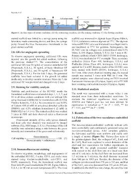
Figure 4. The three types of culture conditions. (A) The obstructing condition. (B) The soaking condition. (C) The flowing condition.
stained scaffold was linked to a syringe pump using the scaffold was immersed in alginate lyase (Sigma-Aldrich,
laboratory made connecting device, and then, the syringe U.S.A.) solution to remove alginate at 37°C. The alginate
pump flowed the blue fluorescence microbeads to the removed HUVEC core was immersed in a collagen matrix
green stained scaffold. and incubated at 37°C for gelation. Subsequently, the
HUVEC core in collagen was permeabilized with 0.1%
2.8. GFs for angiogenic sprouting Triton X-100 (Sigma-Aldrich, U.S.A.) for 5 min at RT.
To observe angiogenic sprouting, additional GFs were Primary antibody of anti-CD31 (MA5-13188, Invitrogen,
injected into the growth kit added medium, following U.S.A.) was incubated at 4°C overnight. Then, secondary
the previous studies [15,21] . The concentration of the antibodies (Alexa Fluor 488, Invitrogen, U.S.A.) and
additional GFs was 50 ng/mL of vascular endothelial GF Phalloidin (Alexa Fluor 488, Invitrogen, U.S.A.) were
(Preprotech, U.S.A.), 50 ng/mL of basic fibroblast GF applied for 2 h at RT. Besides, nuclei of the HUVEC core
(Preprotech, U.S.A.), and 50 ng/mL of hepatocyte GF were stained with DAPI (D1396, Invitrogen, U.S.A.)
(Preprotech, U.S.A.). For the first 2 days, the generated for 5 min. After every chemical treating step, the treated
scaffolds have been cultured in the growth kit added sample was washed 3 times with PBS for 5 min. The
media only to develop vascular structure. Since day 3, the stained samples were observed using an IX53 inverted
additional GF was provided and changed every 2 days. fluorescent microscope (Olympus, Japan) and a FV1000
laser scanning confocal microscope (Olympus, Japan).
2.9. Staining for viability analysis 2.11. Statistical analysis
Viability and proliferation of the HUVEC inside the
formulated scaffold were evaluated at days 1, 3, 5, 7, and The result was represented with a mean value ± one
10 in all three culture conditions with and without GFs standard error from three independent repetitions. To
using live/dead viability kit for mammalian cells (L3224, evaluate the statistical significance level, one-way
Thermo Scientific, U.S.A.). Its concentration was 0.05% ANOVA and Tukey’s post hoc test were utilized. Its
of Calcein AM (4 mM) in anhydrous dimethyl sulfoxide significance is remarked as * for P < 0.05, ** for
(DMSO) and 0.2% ethidium homodimer-1 (2 mM) in P < 0.01, and *** for P < 0.001.
DMSO/H O at 1:4 (v/v). The stained scaffold was washed 3. Results
2
3 times in PBS and then observed under a fluorescent
microscope. 3.1. Fabrication of the two-vasculature-embedded
Fluorescent intensity of live cells (green channel) scaffold
and dead cells (red channel) was analyzed by ImageJ
software (Fiji, NIH Image, U.S.A.). Percentage of the cell The two-vasculature-embedded scaffolds without cells
viability was calculated using a ratio between the green were controllably and continuously generated using our
intensity and summation of the green and red intensity. two-core-embedded device. After complete gelation,
the fabricated scaffolds were uniform and stable with
Green intensity a length of meters (Figure 5A). Various flow rates of
Cell viability = × 100 (1) the gelatin-alginate fluid, the collagen-CaCl fluid, and
Green intensity red intensity+ 2
the CaCl fluid were explored to select diameters of the
2
shell and two vasculatures for further experiments. In
2.10. Immunofluorescent staining Figure 5C, the graph presented scaffolds’ diameter
with respect to the shell flow rate at a fixed core flow
To observe migration, morphology, and angiogenesis of rate of 0.1 mL/min. As the shell flow rate increased from
the embedded HUVEC, F-actin, CD31, and nuclei were 1.5 mL/min to 3 mL/min, the shell diameter increased
stained using rhodamine-phalloidin, anti-CD31, and from 948 µm to 1095 µm. Besides, the diameter of the
DAPI, respectively. First, the formulated scaffold was collagen core decreased from 376 µm to 262 µm, and that
fixed with 4% paraformaldehyde (P6148, Sigma-Aldrich, of the CaCl core also decreased from 331 µm to 203 µm.
2
U.S.A.) for 40 min at room temperature (RT). The fixed In addition to the shell flow rate change, the core flow rate
International Journal of Bioprinting (2022)–Volume 8, Issue 3 57