
Page 215 - IJB-9-1
P. 215
International Journal of Bioprinting FeS /PCL scaffold for bone regeneration
2
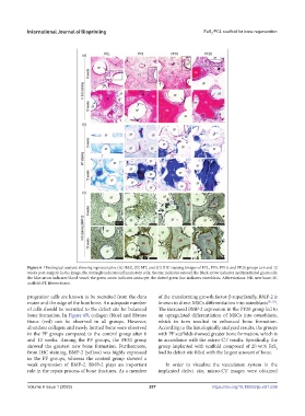
Figure 6. Histological analysis showing representative (A) H&E, (B) MT, and (C) IHC staining images of PCL, PF5, PF10, and PF20 groups at 6 and 12
weeks post-surgery. In the image, the rectangle indicates inflammatory cells; the star indicates osteoid; the black arrow indicates multinucleated giant cells;
the blue arrow indicates blood vessel; the green arrow indicates osteocyte; the dotted green line indicates osteoblasts. Abbreviations: NB, new bone; SF,
scaffold; FT, fibrous tissue.
progenitor cells are known to be recruited from the dura of the transforming growth factor-β superfamily, BMP-2 is
mater and the edge of the host bone. An adequate number known to direct MSCs differentiation into osteoblasts [51,52] .
of cells should be recruited to the defect site for balanced The increased BMP-2 expression in the PF20 group led to
bone formation. In Figure 6B, collagen (blue) and fibrous an upregulated differentiation of MSCs into osteoblasts,
tissue (red) can be observed in all groups. However, which in turn resulted in enhanced bone formation.
abundant collagen and newly formed bone were observed According to the histologically analyzed results, the groups
in the PF groups compared to the control group after 6 with PF scaffolds showed greater bone formation, which is
and 12 weeks. Among the PF groups, the PF20 group in accordance with the micro-CT results. Specifically, the
showed the greatest new bone formation. Furthermore, group implanted with scaffold composed of 20 wt% FeS
2
from IHC staining, BMP-2 (yellow) was highly expressed had its defect site filled with the largest amount of bone.
in the PF groups, whereas the control group showed a
weak expression of BMP-2. BMP-2 plays an important In order to visualize the vasculature system in the
role in the repair process of bone fractures. As a member implanted defect site, micro-CT images were obtained
Volume 9 Issue 1 (2023)olume 9 Issue 1 (2023) 207 https://doi.org/10.18063/ijb.v9i1.636
V