
Page 76 - IJB-9-1
P. 76
International Journal of Bioprinting Osteoconduction and scaffold directionality
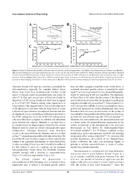
Figure 5. Extent of filament directionality influences osteoconduction and bone regeneration. Correlation of rod dimension/rod distance and direction-
ality on bony bridging (A) and bony regenerated area (B). In (A) and (B), the data set with scaffolds of 100% in direction of bone ingrowth is displayed
2
in black and with 50% directionality in gray. The low R value for directionality of 100% indicates that rod dimension and rod distance have no major
influence on bony bridging (black lines), but at 50% directionality (gray lines), an increase in rod dimension and rod thickness is highly associated with a
decrease in bony bridging. Correlation of total surface area in direction of bone growth on bony bridging (C) and bony regenerated area (D) are shown.
Filaments beyond 0.83 mm are, therefore, suboptimal for from the defect margins toward the center of the defect. A
osteoconduction, especially for complex defects where unilateral vectorized growth pattern is essential for nerve
diverse bone fronts from simultaneously. Another crucial repair and can be generated by a two-photon lithography-
[33]
aspect of filament-based microarchitectures was found by based 3D patterning of 200 nm nanofibers. The dimension
others in that each second layer of filaments should be of these fibers is far below the dimension of the filaments
[23]
aligned in a 0°/90° setting. If only each third layer is aligned used in this study and resembles the situation for guiding the
as in a 0°/60°/120° filament setting, bone regeneration is migration of single cells on a surface . Osteoconduction in
[22]
compromised. This suggests that it is not only the alignment wide-open porous scaffolds, however, is guiding bone tissue
of the filaments of one layer with the bone front to create growth and represents no surface phenomenon since bone
a directional space between two adjacent filaments but also formation does not occur predominantly on the surface of
an alignment of every second layer of filaments provided in the scaffold but between the rods of lattice microarchitectures
the 0°/90° setting but not in the 0°/60°/120° setting where as shown for two different materials: TCP and titanium [1,32] .
only each third layer is aligned. In addition, the directional Therefore, for osteoconduction, the microarchitecture and
space between two adjacent filaments in one layer has to to a lesser extent the nanoarchitecture represented by the
be complemented by aligned spaces formed by the second surface morphology or the microporosity is the dominant
filament layers to create an overall osteoconductive space determinant as was shown for hydroxyapatite-based
configuration. Although directional bone formation 3D-printed scaffolds . For TCP-based scaffolds, surface
[34]
occurs in the space between the filaments and not on their morphology, and/or microporosity tuned by the sintering
surface [1,32] , aligned spaces are defined by aligned surfaces. We temperature affect osteoconductivity . To overcome this
[30]
found that the area of surface aligned to bone ingrowth per possible dependence, all scaffolds of our filament-based
volume is an important determinant of osteoconductivity. library underwent the same post-processing regime to
A value exceeding 0.5 mm per mm of scaffold is sufficient generate a uniform nanoarchitecture in terms of surface
3
2
for this surface to serve as a guiding cue for maximal morphology and microporosity.
osteoconductivity. Values below this threshold, however, Vascularization is a prerequisite for bone formation
correlate strongly with a decrease in osteoconductivity and occurs even in pores between 40 and 70 µm. In a
(Figure 5C) and bony regeneration (Figure 5D).
set of ceramic scaffolds with pores between 0.04 and
The primary criterion for directionality in 0.28 mm, the density of functional capillaries was the
osteoconduction is defect bridging, since in contrast to bony highest with pores exceeding 0.14 mm, and it correlates
regenerated area, it reflects a vectorized growth pattern with the extent of new bone formation [35] . Since the
Volume 9 Issue 1 (2023)olume 9 Issue 1 (2023) 68 https://doi.org/10.18063/ijb.v9i1.626
V