
Page 203 - IJB-9-3
P. 203
International Journal of Bioprinting 3D-printed vascularized biofunctional scaffold
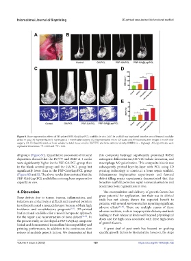
Figure 8. Bone regeneration effects of 3D-printed PRP-GA@Lap/PCL scaffolds in vivo. (A) The scaffold was implanted into the area of femoral condylar
defect in rats. (B) Representative X-ray images at 1 month after surgery. (C) Representative micro-CT scans and 3D reconstruction images 1 month after
surgery. (D, E) Quantification of bone volume to total tissue volume (BV/TV) and bone mineral density (BMD) (n = 6/group). All experiments were
replicated three times. *P < 0.05 and **P < 0.01.
all groups (Figure 8C). Quantitative assessment of mineral this composite hydrogel significantly promoted BMSC
deposition showed that the BV/TV and BMD at 4 weeks osteogenic differentiation, HUVEC tubule formation, and
were significantly higher in the PRP-GA/PCL group than macrophage M2 polarization. This composite bioink was
in the blank control group and the GA/PCL group but subsequently printed layer-by-layer with PCL using 3D
significantly lower than in the PRP-GA@Lap/PCL group printing technology to construct a bone repair scaffold.
(Figure 8D and E). The above results demonstrated that the Subcutaneous implantation experiments and femoral
PRP-GA@Lap/PCL scaffold has a strong bone regenerative defect filling repair experiments demonstrated that this
capacity in vivo. bioactive scaffold promotes rapid neovascularization and
accelerates bone regeneration in vivo.
4. Discussion The encapsulation and delivery of growth factors has
great potential for application, but their use in clinical
Bone defects due to tumor, trauma, inflammation, and trials has not always shown the expected benefit to
infection are collectively a difficult and unsolved problem patients, with several previous studies reporting significant
in orthopedics and a research hotspot because of their high adverse effects [34-36] . There are multiple causes of these
incidence and unsatisfactory prognosis [1,31] . 3D-printed adverse reactions, such as inappropriate delivery methods
biofunctional scaffolds offer a novel therapeutic approach leading to their release at levels well beyond physiological
for the repair and reconstruction of bone defects [32,33] . In doses and the high costs associated with these high doses
the present study, we developed a PRP-GA@Lap composite of growth factors.
bioink and demonstrated its excellent biocompatibility and
printing performance, in addition to its continuous slow A great deal of past work has focused on grafting
release of multiple growth factors. We demonstrated that specific growth factors to biomaterials; however, the steps
Volume 9 Issue 3 (2023) 195 https://doi.org/10.18063/ijb.702