
Page 259 - IJB-9-3
P. 259
International Journal of Bioprinting Multi-material bioprinting with OCT imaging
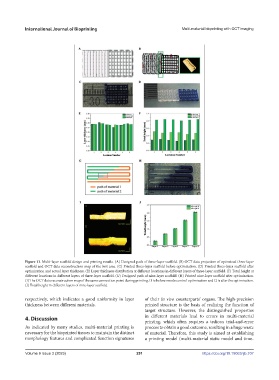
Figure 11. Multi-layer scaffold design and printing results. (A) Designed path of three-layer scaffold. (B) OCT data projection of optimized three-layer
scaffold and OCT data reconstruction map of the box area. (C) Printed three-layer scaffold before optimization. (D) Printed three-layer scaffold after
optimization and actual layer thickness. (E) Layer thickness distribution at different locations in different layers of three-layer scaffold. (F) Total height at
different locations in different layers of three-layer scaffold. (G) Designed path of nine-layer scaffold. (H) Printed nine-layer scaffold after optimization.
(I) The OCT data reconstruction map of the same connection point during printing. I1 is before nozzle control optimization and I2 is after the optimization.
(J) Total height in different layers of nine-layer scaffold.
respectively, which indicates a good uniformity in layer of their in vivo counterparts’ organs. The high-precision
thickness between different materials. printed structure is the basis of realizing the function of
target structure. However, the distinguished properties
4. Discussion in different materials lead to errors in multi-material
printing, which often requires a tedious trial-and-error
As indicated by many studies, multi-material printing is process to obtain a good outcome, resulting in a huge waste
necessary for the bioprinted tissues to maintain the distinct of material. Therefore, this study is aimed at establishing
morphology features and complicated function signatures a printing model (multi-material static model and time-
Volume 9 Issue 3 (2023) 251 https://doi.org/10.18063/ijb.707