
Page 427 - IJB-9-4
P. 427
International Journal of Bioprinting 3D bioprinting of artificial blood vessel
A B
C
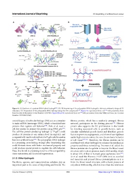
Figure 8. (A) Synthesis of a protein-DNA hybrid hydrogel [146] . (B) 3D bioprinting of the polypeptide-DNA hydrogel to fabricate arbitrarily designed 3D
structures. (C) Preparation of the polypeptide-DNA hydrogel using the two components [147] . Figure 8A reproduced from ref. [146] with permission from
Royal Society of Chemistry (Order Number: 1250780). Figure 8B and 8C reproduced from ref. [147] with permission from John Wiley and Sons, Inc. (License
Number: 5355800472548).
central dogma of molecular biology: DNA acts as a template fibrous protein, which has a randomly arranged fibrous
to make mRNA (messenger RNA), which is translated into network, participates in the clotting process [150] . Fibrous
proteins that regulate cell behavior [148] . Park et al. used a protein also supports the EC proliferation in the bioink
cell-free system to prepare the protein using DNA gels [149] . by directing associated cells to growth factors, such as
The cell-free protein-producing hydrogel (a “P-gel”) could vascular endothelial growth factor and fibroblast growth
produce 16 proteins at very minute level (in mg/mL), and factor, to promote angiogenesis, and shows shear rigidness
compared with traditional method, the P-gel could be used as under high strain to mimic the non-linear elastic behavior
bioink in artificial tissue [149] . The polypeptide-DNA hydrogel of soft tissue [150-152] . Therefore, the fibrous protein can be
is a promising cross-linking strategy after bioprinting that combined with other hydrogels to enhance the mechanical
could fabricate items with better mechanical property and property and tissue remodeling. Freeman et al. added the
could produce special protein to regulate the cell’s behavior. fibrous protein into the gelatin to bioprint the blood vessel
Thus, this bioink is promising in terms of biocompatibility, structure with rupture pressure reach 1110 mmHg, which
tissue maturation, and functional regeneration. is about 52% of that in human’s great saphenous vein [153] .
Li et al. used the Pluronic F-127 (10% w/v) as scarified
3.1.5. Other hydrogels rod materials and printed fibrous protein/gelatin on it to
The fibrin, agarose, and nanocrystalline cellulose play an form the blood vessel structure, with a burst pressure of
important part in the areas of bioprinting and bioink. The only about 1000 mmHg, which is lower than the minimum
Volume 9 Issue 4 (2023) 419 https://doi.org/10.18063/ijb.740