
Page 430 - IJB-9-4
P. 430
International Journal of Bioprinting 3D bioprinting of artificial blood vessel
A
B
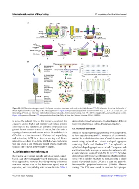
Figure 10. (A) Manufacturing process of 3D alginate container structures with multi-scale fluid channels [174] . (B) Schematic depicting the benefits of
Mitch-Alginate bioink for each stage of the printing process [178] . Figure 10A reprinted (adapted) with permission from “Gao Q, Liu Z, Lin Z, et al., 2017, 3D
Bioprinting of Vessel-like Structures with Multilevel Fluidic Channels. ACS Biomater Sci Eng, 3(3): 399–408.” Copyright 2017 American Chemical Society.
Figure 10B reproduced from ref. [178] with permission from John Wiley & Sons, Inc. (License Number: 5355931318350).
is to use the natural ECM as the bioink to construct the demonstrates the advantages and disadvantages of different
organs to ensure higher cell viability and induce specific bioprinting techniques in blood vessel construction.
cell behavior. The natural ECM contains compounds and
growth factors unique to natural tissues, but also with a 4.1. Material extrusion
topology that compounds cannot mimic. Nonetheless, it is Extrusion-based bioprinting platform is a promising method
difficult to produce the natural ECM required as purifying to form vascular structures [181] . Norotte et al. discovered a
and extracting ECM is a time-consuming and labor- method for scaffold-free fabrication of small-diameter blood
intensive procedure. Despite the ECM shortage, we believe vessels using spheroid or cylindrical-shaped aggregates
that the ECM is the promising bioink which could fully containing SMCs and fibroblasts [182] . The spheroid- or
simulate the complex environment of organs. cylindrical-shaped aggregates were extruded by agarose rods
and then fused to form single- or double-layered vessels with
4. 3D bioprinting techniques an outer diameter ranging from 0.9 to 2.5 mm [182] . Park et al.
Bioprinting approaches include extrusion-based, inkjet- demonstrated extrusion-based bioprinting of artificial blood
based, and stereolithography-based techniques. Among vessel with a tubular structure by manufacturing a single
these approaches, extrusion-based bioprinting is the most strand of polyvinyl alcohol (PVA) as a core and printed a
common method due to fast fabrication speed, ease of biocompatible polydimethylsiloxane (PDMS) filament
operation, and compatibility with various bioinks. Table 3 coating. The PVA core could be removed by hydrogen
Volume 9 Issue 4 (2023) 422 https://doi.org/10.18063/ijb.740