
Page 373 - IJB-9-5
P. 373
International Journal of Bioprinting Multifunctional hydrogel surgical training model
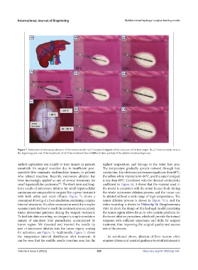
Figure 7. Simulation of microwave ablation of liver tumor model. (a) Conceptual diagram of the structure of the liver organ. (b, c) Cross-sectional view at
the beginning and end of the treatment. (d–i) Cross-sectional view of different time periods of the ablation treatment process.
earliest application was mainly to treat tumors in patients highest temperature and belongs to the water loss area.
unsuitable for surgical resection due to insufficient post- The temperature gradually spreads outward through heat
operative liver remnants, multinodular tumors, or patients conduction. The white area part is more significant than 80°C,
who refused resection. Recently, microwave ablation has the yellow-white interval is 60–80°C, and the outermost part
been increasingly applied as one of several treatments for is less than 60°C. Combined with the thermal conductivity
[35]
small hepatocellular carcinoma . The short-term and long- coefficient in Figure 3d, it shows that the material used in
term results of microwave ablation for small hepatocellular the model is consistent with the actual human body during
carcinoma are comparable to surgery. It is a green treatment the whole microwave ablation process, and the tumor can
with both safety and exact efficacy. Figure 7a shows a be ablated without a wide range of high temperatures. The
conceptual drawing of a liver simulation containing complex tumor ablation process is shown in Figure 7d–i, and the
internal structures. It is often necessary to avoid the complex video recording is shown in Videoclip S6 (Supplementary
systems inside the liver to reach the treatment area accurately File). In short, the design of this hydrogel model containing
under ultrasound guidance during the surgical treatment. the tumor region allows for an in vitro realistic platform for
To facilitate data recording, we designed a simple simulation the tumor ablation procedure, which will provide the trained
sample of simulated liver parenchyma accompanied by surgeons with sufficient experience and skills for surgical
tumor region. We dissected and inserted the needle tip treatment, thus improving the surgical quality and success
part of microwave ablation into the tumor region, waiting rate of the process.
for activation, see Figure 7b. Additionally, Figure 7c shows
the temperature interval distribution after treatment. It As mentioned above, ablation of liver tumors often
can be seen that the middle needle insertion area has the requires ultrasound-assisted guidance to avoid intrahepatic
Volume 9 Issue 5 (2023) 365 https://doi.org/10.18063/ijb.766