
Page 374 - IJB-9-5
P. 374
International Journal of Bioprinting Multifunctional hydrogel surgical training model
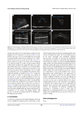
Figure 8. Ultrasonic images of hydrogel models of different organs. (a) Inferior vena cava structure of the hydrogel liver model. (b) Internal tumor of the
hydrogel model. (c) The ablation needle is inserted into the designated tumor site under ultrasound guidance. (d, e) The small and large pancreatic ducts
of the hydrogel pancreas model. (f) The structure of the thyroid gland in the hydrogel roaring neck model. IVC, inferior vena cava.
vessels, veins, and other structures and accurately reach the with biological structures and mechanical properties close
designated site for treatment. As shown in Figure 8a, the to those of natural soft tissues. The mechanical properties
inferior vena cava (IVC) structure inside the liver model of the elastic hydrogels were extensively regulated
is presented under ultrasound at a frequency of 4–6 MHz, and precisely controlled by selecting the appropriate
and the ductal access structure can be seen. Figure 8b–c concentration of monomer composition and immersion
shows ultrasound images of the tumor spheres designed time of the ionic solution. Various functionalized hydrogel
to be detected inside the model and the accurate arrival organ models with structural and mechanical properties
of the ablation needle at the designated tumor area under matching natural soft tissues, including liver, kidney, and
ultrasound, respectively. In addition, we also simulated the blood vessels, were finely fabricated by combining various
ultrasound visualization of the internal structure of the fabrication schemes of 3D printing. The prepared different
pancreas in an actual situation by using an injection needle organ models have physicochemical properties close to
to continuously pass a flowing fluid in the pancreatic duct the actual human body, good feedback from surgical
inside the pancreas, as shown in Figure 8d–e, where the instruments, and realistic haptics and appearance. In
entire interior is visible. To demonstrate the usability of addition, the designed models for hemostasis of blood
the material in various models, in addition to the liver vessels of different diameters, resection and ablation of
and pancreas models, we also tested the prepared thyroid the liver, and ultrasound-assisted visualization of internal
model (Figure S1 in Supplementary File). It was found that structures can be used as practical clinical and surgical
the test results still matched the actual human tissue and training platforms for in vitro simulation and training.
were consistent with the expected objectives, as shown in In summary, the solution based on the combination of
Figure 8f. In other words, the visualization of the internal hydrogel tissue simulants with mechanically adjustable
structure of the hydrogel model can be achieved through softness and advanced 3D printing manufacturing
the differentiated design of the matrix part and the internal technology is considered to have a wide range of
construction of the model. This design solution will applications as a deployable platform for in vitro demand
provide a novel and convenient platform for training and testing of new medical devices, preoperative rehearsal, and
teaching many procedures. surgical training.
4. Conclusion
In conclusion, we propose novel elastic hydrogels with tissue Acknowledgments
softness suitable for simulating tissue and organ models None.
Volume 9 Issue 5 (2023) 366 https://doi.org/10.18063/ijb.766