
Page 386 - IJB-9-5
P. 386
International Journal of Bioprinting Vascularized bone regeneration
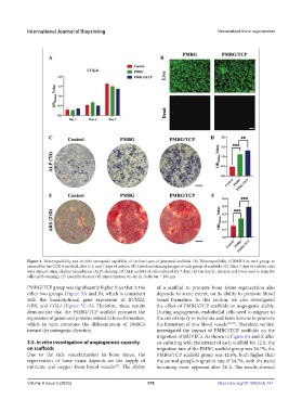
Figure 4. Biocompatibility and in vitro osteogenic capability of various types of prepared scaffolds. (A) Biocompatibility of BMSCs in each group, as
assessed by the CCK-8 method, after 1, 4, and 7 days of culture. (B) Live/dead staining images of each group of scaffolds. (C) After 7 days of culture, cells
were stained using alkaline phosphatase (ALP) staining. (D) ALP activity of cells cultured for 7 days. (E) On day 21, Alizarin red S was used to stain the
cells (ARS staining). (F) Quantification of cell mineralization on day 21. Scale bar = 200 μm.
PMBG/TCP group was significantly higher than that in the of a scaffold to promote bone tissue regeneration also
other two groups (Figure 5A and B), which is consistent depends, to some extent, on its ability to promote blood
with the transcriptional gene expression of RUNX2, vessel formation. In this section, we also investigated
OPN, and COL1 (Figure 5C–E). Therefore, these results the effect of PMBG/TCP scaffolds on angiogenic ability.
demonstrate that the PMBG/TCP scaffold promotes the During angiogenesis, endothelial cells need to migrate to
expression of genes and proteins related to bone formation, the site of injury or ischemia and form lumens to promote
which in turn promotes the differentiation of BMSCs the formation of new blood vessels [38,39] . Therefore, we first
toward the osteogenic direction. investigated the impact of PMBG/TCP scaffolds on the
migration of HUVECs. As shown in Figure 6A and B, after
3.5. In vitro investigation of angiogenesis capacity co-culturing with the extract of each scaffold for 12 h, the
on scaffolds migration rate of the PMBG scaffold group was 34.7%, the
Due to the rich vascularization in bone tissue, the PMBG/TCP scaffold group was 42.6%, both higher than
regeneration of bone tissue depends on the supply of the control group’s migration rate of 34.7%, with the trend
nutrients and oxygen from blood vessels . The ability becoming more apparent after 24 h. The results showed
[37]
Volume 9 Issue 5 (2023) 378 https://doi.org/10.18063/ijb.767