
Page 391 - IJB-9-5
P. 391
International Journal of Bioprinting Vascularized bone regeneration
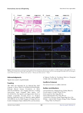
Figure 9. Histomorphometric assessment of bone regeneration at specific time intervals for each Implant. (A) H&E staining for each implantation in vivo.
(B) Masson’s trichrome staining for each implantation in vivo. (C) Immunofluorescence images of in vivo tissue angiogenesis marker CD31 and osteogenic
marker OCN. Red scale bar = 100 µm, black scale bar = 1000 µm, and white scale bar = 100 µm.
Acknowledgments of National Facility for Translation Medicine (Shanghai)
(TMSK-2020-118, TMSK-2021-140).
Figures were created using BioRender.
Conflict of interest
Funding
The authors declare no conflict of interest.
This study was supported by the National Key R&D
Program of China (2022YFA1207500/2018YFA0703000), Author contributions
National Natural Science Foundation of China
(82072412/92048205), 2022 Lingang laboratory “Seeking Conceptualization: Changru Zhang, Ya Ren, Heyue Li
Outstanding Youth Program” open project (LG- Formal analysis: Changru Zhang, Ya Ren,
QS-202206-04), Biomaterials and Regenerative Medicine Fund acquisition: Han Yang, Bin Cai
Institute Cooperative Research Project by Shanghai Project administration: Han Yang, Bin Cai
JiaoTong University School of Medicine (grant number Visualization: Chengwei Wang, Liang Tang, Haoyi Niu
2022LHBO8), the Key R&D Program of Jiangsu Province Writing – original draft: Changru Zhang, Ya Ren, Weiqing
Social Development Project (BE2022708), and Foundation Kong, Yihao Liu, Heyue Li
Volume 9 Issue 5 (2023) 383 https://doi.org/10.18063/ijb.767