
Page 387 - IJB-9-5
P. 387
International Journal of Bioprinting Vascularized bone regeneration
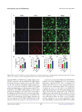
Figure 5. Effects of prepared scaffolds on osteogenic differentiation. (A, B) Immunofluorescence confocal images and quantitative analysis of the osteogenic
markers COL1 (scale bar =100 μm). Osteogenic gene expression of (C) OPN, (D) COL1, and (E) RUNX2.
that both PMBG and PMBG/TCP scaffolds could promote Furthermore, we also explored the ability of PMBG/TCP
HUVEC migration, with the PMBG/TCP scaffold group scaffolds to promote angiogenesis at the transcriptional
having the strongest migration-promoting effect. Based and protein expression levels. After co-culturing each
on the tube formation experiment results presented in scaffold with HUVECs for 2 days, immunofluorescence
Figure 6C, it was observed that the PMBG/TCP scaffold experiments were performed to detect CD31 expression.
group had a higher degree of tube formation with HUVECs As shown in Figure 7A, HUVECs in the PMBG/TCP
compared to the PMBG scaffold group and the control scaffold group expressed more CD31 (green fluorescence),
group at the designated time point. After using Image J and in the fluorescence quantification, the PMBG/TCP
to calculate the results of the tube formation experiment, scaffold group also showed the best ability to promote
we found that the various parameters calculated for the CD31 expression (Figure 7B). Real-time PCR experiments
PMBG/TCP scaffold group were all higher than those for were performed to detect the ability of each scaffold to
the control group and the PMBG scaffold group, and all promote angiogenesis at the gene transcription level.
showed a trend of increasing over time (Figure 6D and E). The study findings revealed that the PMBG/TCP scaffold
Volume 9 Issue 5 (2023) 379 https://doi.org/10.18063/ijb.767