
Page 389 - IJB-9-5
P. 389
International Journal of Bioprinting Vascularized bone regeneration
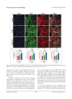
Figure 7. Effects of various prepared scaffolds on angiogenesis. (A, B) Immunofluorescence confocal images and quantitative analysis of the angiogenesis
markers CD31 (scale bar = 50 μm). Angiogenesis gene expression of (C) CD31, (D) FGF, and (E) ANG.
points (Figure 8C and D). These results indicate that the with scattered fibrous tissue. The PMBG/TCP scaffold
PMBG/TCP scaffold successfully repaired critical-sized group, due to the addition of TCP, showed superior bone
bone defects, and the PMBG/TCP scaffold group had a regeneration ability compared to the PMBG scaffold
faster and better new bone fusion rate compared to the group. Additionally, it could be observed that the PMBG/
other groups. TCP scaffold filled the defect, and the surface pores of
the scaffold were filled with newly formed mineralized
Furthermore, H&E and Masson’s trichrome staining
were applied to investigate the effect of the prepared bone. The newly formed bone tissue filled all corners of
the scaffold and even extended to the entire defect area.
scaffolds on bone regeneration. As shown in Figure 9A Furthermore, more bone tissue growth promoted scaffold
and B, no inflammation or necrosis was observed in the degradation.
stained sections of all groups after implantation. Moreover,
new bone and fibrous tissue appeared in the PMBG It has been reported that silicon elements can
scaffold group and the PMBG/TCP scaffold group, while promote vascular regeneration, which is crucial for bone
the defect area in the control group was only partially filled regeneration [40,41] . Therefore, we further validated the ability
Volume 9 Issue 5 (2023) 381 https://doi.org/10.18063/ijb.767