
Page 388 - IJB-9-5
P. 388
International Journal of Bioprinting Vascularized bone regeneration
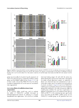
Figure 6. Capability of prepared scaffolds to promote migration and tube formation of HUVECs. (A) After co-culturing various groups of scaffolds with
cells for 12 and 24 h, cell migration images were observed. (B) Analysis of cell migration rate using ImageJ. (C) Formation of endothelial network by
HUVECs after 3 and 6 h of cell culture. (D, E ) The number of junctions (D) and total length (E) per high power field (HPF) were analyzed. Scale bar
=200 μm.
group was more effective in promoting the expression of implanted during surgery fit well with the defect site,
ANG, FGF, and CD31 genes, compared to both the control demonstrating their personalized fabrication. Throughout
group and the PMBG scaffold group (Figure 7C–E). All the entire animal experiment period, no infections or
of these results indicate that PMBG/TCP has the ability deaths were observed. Rats were euthanized at 6 and 12
to promote angiogenesis and can be used for vascularized weeks after surgery, and X-ray analysis was performed on
bone regeneration. the skulls. As shown in Figure 8A and B, the PMBG/TCP
scaffold group significantly enhanced new bone formation
3.6. In vivo effects of scaffold on bone tissue and growth compared to the other two groups, while the
regeneration control group exhibited the least bone regeneration. In
A rat calvarial defect model was used to evaluate addition, quantitative micro-CT analysis showed that the
the ability of PMBG/TCP scaffolds fabricated by BMD and BV/TV in the control group were significantly
photopolymerization-assisted 3D printing to promote lower than those in the PMBG scaffold group and the
vascularized bone regeneration. The cylindrical scaffolds PMBG/TCP scaffold group at all predetermined time
Volume 9 Issue 5 (2023) 380 https://doi.org/10.18063/ijb.767