
Page 113 - IJB-9-6
P. 113
International Journal of Bioprinting Affordable temperature-controlled bioprinter
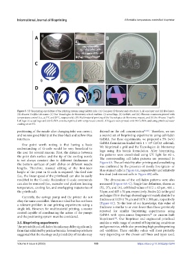
Figure 5. 3D bioprinting capabilities of the printing system using GelMA inks. (A) Complex 3D honeycomb structure: (i, ii) one layer and (iii) five layers.
(B) Alvarez-Trujillo Lab name. (C) The Tecnologico de Monterrey school emblem: (i) actual logo, (ii) GelMA, and (iii) Pluronic constructs printed with
temperature control (i.e., at 5°C and 35°C, respectively). (D) Multimaterial printing of the Tecnologico de Monterrey mascot, and (E) the Alvarez-Trujillo
Lab logo: (i) actual logo and (ii) GelMA construct printed with temperature control. All figures were printed with 5% GelMA; and using printhead water
cooling set at 0°C.
positioning of the nozzle after changing inks was correct, depend on the cell concentration [41,42] . Therefore, we ran
and we saw good fidelity at the blue-black and yellow-blue a second set of bioprinting experiments using cell-laden
interfaces. GelMA. For these experiments, we prepared a 5% (w/v)
6
One point worth noting is that having a basic GelMA formulation loaded with 1 × 10 C2C12 cells/mL.
understanding of G-code would be very beneficial to We bioprinted a grid and the Tecnologico de Monterrey
the user for several reasons. First, the distance between logo using this bioink formulation. After bioprinting,
the petri dish surface and the tip of the cooling nozzle the patterns were crosslinked using UV light for 60 s.
is not always constant due to different thicknesses of The corresponding cell-laden patterns are presented in
the bottom surfaces of petri dishes or different needle Figure 6A. The cell viability after printing and crosslinking
lengths. Therefore, manual editing of the first-layer was confirmed by the presence of mostly live (green- or
height of the print in G-code is required. The feed rate blue-stained cells in Figure 6A, respectively) and relatively
(i.e., the linear speed of the printhead) can also be easily few dead (red-stained cells in Figure 6B) cells.
modified in the G-code. Redundant G-code commands The dimensions of the cell-laden patterns were also
can also be removed (i.e., extruder and platform heating measured (Figure 6A–C). Single-line definition, shown as
temperature, cooling fan, and overlapping trajectories of (X), (Y), and (A), exhibited values of 612 ± 63 µm, 466 ±
the printhead). 51 µm, and 485 ± 51 µm, respectively. Nodes (Z) in the grid
Currently, the syringe pump and the printer do not and edges (B) in the logo showed approximately double the
obey the same controller. This is not ideal but has not been thickness at 1128 ± 76 µm and 1176 ± 308 µm, respectively
a relevant problem in our printing experiments using a (Figure 6C). To the best of our knowledge, this value of
single ink. However, for multimaterial printing, a master thickness is similar to or even lower than those previously
control capable of coordinating the action of the pumps reported for similar bioprinting experiments using
[2]
and the positioning system must be considered. GelMA with open-source bioprinters or custom-built
[34]
bioprinters . Our bioprinter and engineered printhead
3.5. Bioprinting experiments enables a wide range of control of bioprinting conditions
The printability of cell-laden bioinks may differ significantly and geometries, while also providing high postbioprinting
from that exhibited by pristine bioinks. Several reports have cell viabilities. These viability values will most probably
suggested that the rheology and printability of bioinks may vary depending on the chosen cell lines and types used.
Volume 9 Issue 6 (2023) 105 https://doi.org/10.36922/ijb.0244