
Page 450 - IJB-9-6
P. 450
International Journal of Bioprinting 3D bioprinting for lung tissue
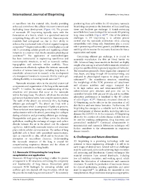
or nanofibers into the material inks, thereby providing positioning these cells within the 3D structure, nanoscale
enhanced control over the cellular microenvironment and bioprinting can promote the formation of functional lung
facilitating tissue development (Figure 4A). The process tissue and facilitate gas exchange. A bioink-containing
of nanoscale 3D bioprinting typically starts with the nanofibrils have been developed for nanoscale 3D-printing
[80]
formulation of a bioink, which is a specialized material lung tissue scaffolds (Figure 4B) . One of the primary
comprising living cells and biomaterials. Nanocomposite challenges in 3D bioprinting is to achieve precise
materials find widespread use due to their ability to control over the nanoscale architecture while ensuring
enhance the mechanical traits of hybrid organic/inorganic compatibility with living cells. Nanoforms play a crucial
[74]
composites . Engineered nanofiber networks play a crucial role in promoting cell survival, growth, and differentiation,
role in promoting cellular growth and regulating cellular enabling cells to assume the necessary functions for tissue
behaviors in a manner that closely emulates physiological regeneration and repair.
conditions . The application of nanotechnologies Concerning efficient gas exchange, it is crucial to
[75]
has been instrumental in engineering nanofibrous and accurately manufacture the thin air–blood barrier via
nanocomposite structures, as well as nanoscale surface LTE. Advanced lung tissue models in the field are highly
topographies and networks within scaffolds. These sought-after, aiming to achieve both biomimetic structural
advancements effectively replicate the intricate nanoscale properties and the ability to precisely regulate cell behavior.
structure of various tissue types, including lung tissue. A The researchers prepared a three-organ chip composed of
remarkable advancement in research is the development liver, heart, and lung through 3D nanobioprinting, and
of a transparent biomimetic nanoscale fibrillar matrix gel, evaluated its physiological response to drugs and toxic
offering flexibility in choosing bioink materials . substances . The nanofibrous structure, resembling
[76]
[81]
Nanoscale structure refers to the detailed anatomical the morphology of the ECM, promotes cell attachment
components and organization of the lung at the nanoscale and enhances nutrition and oxygen transport due
[82]
level . It involves the study and understanding of the to its high surface area and interconnectivity . The
[77]
structures and processes that occur at the nanoscale submicrometer pore structure and pore size can be
within the lung tissue. The alveoli, which are the smallest controlled between 1000 µm and 10 nm, and its excellent
pulmonary functional units, have complex nanostructures. adsorption performance is beneficial to the 3D culture
[83]
The walls of the alveoli are extremely thin, facilitating of cells (Figure 4C) . Nanoscale structural elements in
efficient gas exchange . The alveoli are lined with a 3D bioprinting can be effective in the promotion of cell
[78]
surfactant layer, which is composed of lipids and proteins. distribution and new tissue formation. Furthermore, 3D
These surfactant monolayers play a critical role in reducing bioprinting has emerged as a valuable tool in the design
surface tension within the alveoli, preventing their collapse and development of disease models, including infectious
diseases like COVID-19. At the nanoscale, 3D bioprinting
during exhalation and promoting efficient gas exchange. allows for the creation of realistic disease models that can
Nanoparticles and gases can diffuse across the alveolar be used for studying pathogenesis, drug discovery, and
epithelium, enabling the exchange of oxygen and carbon personalized medicine . Continued advancements in
[84]
dioxide between the air in the alveoli and the adjacent nanomaterials and nanoscale fabrication techniques will
capillaries. Additionally, nanoscale vesicles and exosomes further contribute to the advancements in respiratory
play a role in cellular communication. The surface of lung disease research.
epithelial cells is lined with specialized nanostructures,
such as microvilli or cilia, which aid in functions like 6. Challenges and future directions
absorption or mucociliary clearance. Understanding
the nanoscale structure of the lung is crucial for 3D bioprinting is a promising technology with vast
comprehending respiratory diseases and the effects of potential in tissue engineering, although it is still in its
nanoscale interactions on lung health. early stages of development. Several technical challenges
must be addressed, particularly achieving high-resolution
5.2. Nanoscale 3D bioprinting for lung tissue cell patterning and distribution. Current techniques like
recapitulation and application material extrusion have several drawbacks, including
Nanoscale 3D bioprinting technology enables the precise low cell viability, resolution, and working speed, which
arrangement in 3D structures, mimicking the complexity hinder the fabrication of submicroscale and nanoscale
and functionality of lung tissues. Researchers print structures. To enable the production of macroscale tissues
nanoscale ECM hydrogels by extruding cellular and for clinical applications, printing capabilities and speed
acellular gels into stacked cell ring structures, which has must be improved . In addition, for successful in vivo
[85]
the potential to study lung nanostructures . By accurately transplantation, the printed tissue must possess appropriate
[79]
Volume 9 Issue 6 (2023) 442 https://doi.org/10.36922/ijb.1166