
Page 541 - IJB-9-6
P. 541
International Journal of Bioprinting High-performance SrCS scaffolds via vat photopolymerization
exhibited a limited number of micropores, suggesting a about three times that of the pure scaffold. One of the
lower degradation rate. reasons is that composite scaffolds lost components
Particularly, the degradation rate of the SrCS-40BTA more slowly in the degradation process, resulting in
scaffold after soaking for 14 days was only 1.32 ± 0.09%, more complete structures and fewer surface defects
which was one-third that of the pure SrCS scaffold (shown than the pure SrCS scaffold. Another reason is that the
in Figure 11a). Accordingly, as the structure was relatively pinning effect played a crucial role in hindering the crack
complete, the compressive strength of the composite extension and grain movement during the compression
scaffolds was also better than the pure SrCS scaffold after process, and became more obvious with the increase
degradation (shown in Figure 11b). Moreover, the SrCS- of BTA content. Meanwhile, the elasticity modulus
40BTA scaffold exhibited a few microcracks after soaking of the scaffolds increased after in vitro degradation
for 4 days but was intercepted by the CaTiO grains. (shown in Figure 12d). According to previous reports,
3
This pinning effect was one of the main reasons for the an elastic modulus greater than 3.0 GPa would not
[55]
enhancement of mechanical properties, in consistent be conducive to the growth of new bone . Thus, we
with the previous results. should adjust the content of BTA so that the composite
scaffolds have the required compressive strength while
The degraded scaffolds were subjected to compression maintaining the appropriate elasticity modulus. In vitro
tests to evaluate their mechanical properties in operation degradation experiment revealed that we can regulate
(shown in Figure 12a). Figure 12b presents the stress– the biodegradability of SrCS scaffolds by changing the
strain curves of the scaffolds after in vitro degradation content of BTA to provide the necessary mechanical
for 14 days, and the curves of other days are shown in support for the long-time regeneration of large
Figure S4 (Supplementary File). These curves all showed bone defects.
that scaffolds doped with BTA still had high compressive
strength after in vitro degradation, which ensured that 3.5. In vitro biological activity evaluation
they could play a good supporting role in the process of To verify the biological activity of the scaffolds, we tested
bone repair. In addition, the greater energy absorption of the OD values of cell proliferation by CCK-8 assay, as
the composite scaffolds resulted in a strong deformation shown in Figure 13. After culturing for 1 day, the OD values
resistance. Figure 12c shows the compressive strength of SrCS composite scaffolds were similar to those of the
of the degraded scaffolds. The compressive strength of control group. After culturing for 7 days, all components
the SrCS-40BTA scaffold after in vitro degradation was of the cells had obvious proliferation effect, and the SrCS
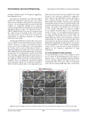
Figure 10. Surface morphology of bioceramic scaffolds with different SrCS-BTA components after soaking for 0, 4, 7, and 14 days, respectively.
Volume 9 Issue 6 (2023) 533 https://doi.org/10.36922/ijb.1233