
Page 71 - IJB-9-6
P. 71
International Journal of Bioprinting 3D Aerosol Jet® printing for microstructuring
3D AJ P results on AgNPs-based ink
®
Not diluted Diluted (AgNPs:DI water ratio of 1:4)
a b 1 b 2 b 3 b 4
CJD
100 µm 150 µm 150 µm 150 µm 150 µm
c c d 1 d 2 d 3 d 4
LBL
500 µm 250 µm 100 µm 50 µm 50 µm
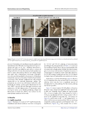
Figure 2. Results for the 3D AJ®P of microstructures for AgNPs-based ink, along with print strategy and ink dilution. (a) Nondiluted and (b ) diluted
1-4
3D-CJD AgNPs-based ink; (c) nondiluted and (d ) diluted 3D-LBL AgNPs-based ink.
1-4
protocol. Particularly, after medium removal, samples were for 3D-CJD and 3D-LBL printing of microstructures
washed twice with PBS and a staining solution (0.5 µL mL according to the print parameters of Table 2. It is observed
−1
calcein-AM and 2.0 µL mL ethidium homodimer-1 that nondiluted AgNPs-based ink generated dendritic-like
−1
[EthD-1] in PBS) was added to each well at a volume of structures with a rough surface. Instead, the diluted version
200 µL/well. After 30 min of incubation, fresh medium was gave rise to denser and smoother 3D printed structures. A
used to replace the staining solution and cellular images certain degree of complexity is also achievable. Moreover,
were taken with a fluorescence microscope (Olympus). the 3D-LBL strategy is preferred over than 3D-CJD due to
Live cells were distinguished by the presence of ubiquitous the higher degree of repeatability and control over printing.
intracellular esterase activity, determined by the enzymatic With respect to the 3D-CJD strategy, Figure 2a shows a
conversion of the virtually nonfluorescent cell-permeant 3D-CJD dendritic-like pillar achieved with the nondiluted
calcein-AM to the intensely fluorescent calcein. The solution, which can also have a significant amount of
polyanionic dye calcein was well retained within live cells, overspray due to a dry printing. For this reason, printing
producing an intense, uniform green fluorescence in live was barely repeatable.
cells. EthD-1 entered cells with damaged membranes and
underwent a 40-fold enhancement of fluorescence upon Figure 2b instead depicts 3D-CJD pillars or branches
binding to nucleic acids, thereby producing a bright red obtained using a diluted ink (AgNPs:DI water ratio of 1:4).
fluorescence in dead cells. EthD-1 was excluded by the In this case, vertically aligned pillars are approximately
intact plasma membrane of live cells. 590 ± 42 µm in height, with a base diameter of 92.95 ±
11.98 µm, a tip diameter of 54.64 ± 5.04 µm, and a 1<AR<12
3. Results based on the applied focusing ratio (R ) . For instance, in
f
Figure 2b , pillars with an AR = 1.7 are obtained at R = 4,
1
f
3.1. AgNPs-based ink with a medium process reproducibility. With an increasing
Figure 2 illustrates the results of AJ®P AgNPs-based inks. complexity printing approach, poly-branches (duo-, tri-,
Nondiluted and diluted solutions were both investigated multibranch, Figures 2b ) were created from a mono-
2-4
Volume 9 Issue 6 (2023) 63 https://doi.org/10.36922/ijb.0257