
Page 40 - TD-3-1
P. 40
Tumor Discovery Energy metabolism in bladder cancer
which, in turn, were blocked with 3% BSA solution diluted pTa tumors was 3.0 cm, and the one for high-grade pT1
in TBS-T buffer to mitigate non-specific protein binding. tumors was 4.0 cm (Table 1).
Nitrocellulose membranes were incubated overnight with Each sample collected from urinary bladders was
primary antibodies (diluted 1:1000 in 1% BSA), such as classified based on their histopathological grade. After the
GLUT1, PFK, GAPDH, LDH, PDH, CS, HADHSC, β-F1- classification procedure, the samples were divided into
ATP synthase (ATPase), and hsp60, at 4°C. On completing four groups based on histopathological grade, namely,
the primary antibody incubation, membranes were further normal (no urothelial lesions) group, low-grade pTa group,
incubated with secondary HRP-conjugated antibodies high-grade pTa group, and high-grade pT1 group. Samples
(diluted 1:3000 ratio in 1% BSA; MilliporeSigma, USA) belonging to the low-grade pTa group showed extensive
for 2 h. Immunoreactive bands were visualized through papillary lesions. Urothelial cells presented overall orderly
incubation with 3,3’-diaminobenzidine chromogen appearance with minimal variability in their architecture
(Sigma Chemical Co., St Louis, USA). Immunoblots were and cytological features, lack of nuclear hyperchromasia,
run in duplicate, and the samples were grouped into sets and infrequent miotic figures (Figure 1A and B). High-
comprising 10 samples per group, for each repetition. grade pTa group samples also presented extensive papillary
Semi-quantitative densitometry analysis was applied to lesions but featured disorderly arranged urothelial cells,
the bands in NIH ImageJ 1.47v software (National Institute significant cell pleomorphism, nuclear hyperchromasia,
of Health, USA, available at: http://rsb.info.nih.gov/ij/), and several mitotic figures (Figure 1C and D). On
and it was followed by statistical analyses. Results are the other hand, the high-grade pT1 group samples
expressed as mean ± standard deviation of band intensities presented basement membrane rupture with consequent
in comparison to β-actin (which was used as endogenous invasion of neoplastic urothelial cells arranged in cords
positive control) labeling intensity. 21 or nests on the lamina propria. Neoplastic urothelial
cells presented eosinophilic cytoplasm and a large
2.4. IBEC number of hyperchromatic nuclei and mitotic figures
The IBEC was calculated from several parameters (Figure 1E and F).
determined based on the established protocols using 3.2. The oxidative phosphorylation pathway
Equation 1. 17,18,23 prevailed in normal bladder tissue and low-grade
2.5. Statistical analysis pTa bladder tumor
Quantitative results are expressed as mean ± standard Intense immunoreactivity was observed for β-F1-ATP
deviation, whenever appropriate. Comparison of synthase (ATPase), HADHSC, PDH, and CS, within the
immunohistochemical and Western blotting data
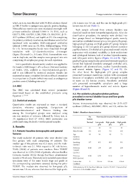
among the investigated groups were analyzed using Table 1. Baseline characteristics
one-way analysis of variance, followed by Tukey test, at No Non‑muscle‑invasive bladder cancer
1% significance level (P <0.01). IBEC assessment was lesions
performed using Student’s t-test for paired samples. Normal Low‑grade High‑grade High‑grade
pTa pTa pT1
3. Results Patients (n) 10 10 10 10
3.1. Patients’ baseline demographic and general Male/Female 7/3 8/2 7/3 8/2
features Age 61 56 62 71
The study included 40 patients who were divided into (years in
median)
four groups, with each comprising 10 patients. The
median age of the patients was 62.5 years, and the male- Smoking 3/7 8/2 8/2 9/1
(yes/no)
to-female ratio was 2:1. With respect to BC risk factors, Previous 0/10 1/9 2/8 2/8
70.0% (28/40) of patients were smokers. Prior TURBT TURBT
was observed in 12.5% (5/40) of patients, whereas Bacillus (yes/no)
Calmette–Guérin (BCG) therapy had been administered Previous 0/10 0/10 0/10 1/9
in only 2.5% of cases. This study only included cases whose BCG (yes/no)
specimens were of the same tumor stage and grade as Lesion size 0 3.0 cm 3.0 cm 4.0 cm
the corresponding specimens assessed during the prior (median)
TURBT. The median size observed for low-grade pTa Abbreviations: BCG: Bacillus Calmette–Guérin; TURBT: Transurethral
tumors was 3.0 cm, whereas that observed for high-grade resection of the bladder tumor.
Volume 3 Issue 1 (2024) 4 https://doi.org/10.36922/td.2290