
Page 207 - IJB-9-5
P. 207
International Journal of Bioprinting Using droplet jetting for bioprinting
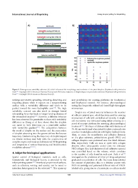
Figure 5. Heterogeneous wettability substrate (A) which influenced the morphology and resolution of ink-jetted droplets (Reprinted with permission
from [109] . Copyright (2014) American Chemical Society) and (B) bounce trajectory of droplet impact on penetrative substrate (Reprinted with permission
from [113] . Copyright (2016) American Chemical Society).
undergo asymmetric spreading, retracting, detaching, and new possibilities for creating platforms for biochemical
migrating phases when it impacts on a nonpenetrating and biophysical research. For instance, pharmacological
surface with a wettability difference and tends to be testing has frequently utilized cell-based high-throughput
pushed toward the more hydrophilic area [118] . The high microarrays.
wettability contrast can also result in stronger lateral Droplet size of jetted material influences the number
rebounding force that leads to longer landing distance of of cells per printed spot, which has been used for creating
the rebounded droplets [119] . However, a different behavior microarrays of cells with controlled cell density. A single-
has been observed for penetrable surfaces with wettability cell microarray was fabricated using inkjet printing as a
difference as Zhang et al. have shown that the droplets proof-of-concept platform for assessing pharmacological
can move in both directions on a penetrable surface treatment [123] . Thin layer of chitosan film with thickness of
with wettability gradient [113] . The competition between 70–80 nm was formed when jetted droplets coalesced onto
the recoil of droplet on the surface and the penetration a surface-treated glass substrate with higher hydrophilicity.
of droplet advancing into the groove defines the bounce Then, to create the nonadhesive and adhesive domains
trajectory. Understanding the trajectory of droplet impact on the glass substrate, poly(ethylene glycol) (PEG) and
on diverse surfaces may help with the construction of collagen droplets were patterned onto the dried chitosan
complex engineered tissue as multimaterial 3D bioprinting film, respectively. Cells are seen at spots with collagen
and integration of various bioprinting and biofabrication droplets when subsequently seeded onto the chitosan/
modalities advance [120,121] .
PEG/collagen film. Spot diameter of cell-adhesive material
4. Inkjet for biological applications was controlled based on the volume, which correlates
to the number of cells found in each spot. Park et al.
[58]
Spatial control of biological materials, such as cells, leveraged on the pixelation of inkjet printing and printed
biomaterials, and biological factors, is provided by the graduated concentration of cells. The team demonstrated
inkjet printing system [122] . Interfacial properties of surfaces, the concept of pixelation akin to a desktop printer with
such as adhesion, wetting, and opacity, can be tuned to RGB ink cartridge and patterned different ratio of 3 RGB-
introduce heterogeneity to the substrate . This opens up labeled cells, giving rise to a heterotypic co-culture model
[79]
Volume 9 Issue 5 (2023) 199 https://doi.org/10.18063/ijb.758